Circulatory System -Heart
Q. Identify and give functions for each of the following: Left and Right Atria, Left and Right Ventricles, Coronary Arteries and Veins, Anterior and Posterior Vena Cava, Aorta, Pulmonary Arteries and Veins, Pulmonary Trunk, Atrioventricular Valves, Chordae Tendinae, Semi Lunar Valves, Septum.
A. The right atrium is the first chamber and sends blood to the right ventricle. The left atrium is the third chamber and sends blood to the left ventricle. The right ventricle is the second chamber and sends blood to the lungs through the pulmonary arteries. The left ventricle is the fourth chamber and sends blood to the body through the aorta. The Coronary arteries supply the heart with blood while the Coronary veins take that blood away. The Anterior Vena Cava takes blood to the heart from the head, the Posterior Vena Cava takes blood from the legs to heart. The aorta receives blood and sends it to the rest of the body. The Pulmonary arteries take blood from the heart to the lungs, the Pulmonary veins take blood from the lungs to the heart. The Pulmonary Trunk is the place just before the artery splits into two and heads for the lungs. The Atrioventricular Valves keep the blood from flowing from the ventricles and back into the atria. The Chordae Tendinae are strings that keep the AV Valves shut. The Semi Lunar Valves keep blood from flowing from the Pulmonary artery and aorta and into the ventricles. The Septum separates the left and right sides of the heart.
Q. Describe the locations and functions of the SA node, AV node, and Purkinje fibres.
A. The SA and AV nodes are located in the right atrium. The Purkinje fibres are in the muscle around the ventricles. The SA nodes cause the atria to contract and pump blood into the ventricles. The AV node sends a signal to the Purkinje fibres which cause the ventricles to contract and pump blood into the pulmonary artery/ aorta.
Q. Describe the autonomic regulation of the heartbeat by the nervous system.
A. When blood pressure is low, the autonomic nervous system causes the heart to beat faster and constricts blood vessels. When the pressure is high, it causes the heart to beat slower and relaxes blood vessels.
Q. Relate factors that affect and regulate blood pressure to hypertension and hypotension.
A. Hypertension is high blood pressure that is caused by: hardening of arteries, diet, obesity, and hormone malfunction. Hypotension is low blood pressure caused by weak pumping of the heart.
Q. Demonstrate the measurement of blood pressure.
A. A blood pressure of 115/75 is normal. The 115 is the systole measurement and range from 110 to 150. The 75 is the diastolic measurement and will be 90 or less.
Q. Distinguish between systolic and diastolic blood pressure.
A. Systolic is when the heart is pumping. Diastolic is when the heart, specifically the ventricles, is relaxing.
Circulatory System - Circulation and Blood
Q. Describe and differentiate among the five types of blood vessels.
A. Arteries have thick walls with three layers. They carry blood from the heart to the arterioles. Arterioles also have thick walls with three layers, but they are smaller than arteries. They carry blood from the arteries to the capillaries. Capillaries are tiny and have thin, one-cell thick walls. They carry blood to cells throughout the body and exchange gases, and nutrients. Venules have thin walls with three layers. They carry blood from capillaries to Veins. Veins also have thin walls with three layers, but they are larger than venules.
Q. Identify and give functions for each of the following: Subclavian Arteries and Veins, Jugular Veins, Carotid Arteries, Anterior and Posterior Vena Cava, Pulmonary Veins and Arteries, Hepatic Vein, Hepatic Portal Vein, Renal Arteries and Renal Veins, Iliac Arteries and Veins, Coronary Arteries and Veins, Aorta.
A. The Subclavian Arteries carry blood to the arms, Subclavian Veins take blood away from arms. Jugular Veins take blood from the head to the Anterior Vena Cava. Carotid Arteries carry blood from the Aorta to the head. The Anterior Vena Cava takes blood from the upper body and takes it to the heart. The Posterior Vena Cava takes blood from the lower body and takes it to the heart. Pulmonary Veins carry deoxgenated blood from the heart to the lungs to oxygenize it. Pulmonary Veins take oxygenated blood from the lungs to the heart. The Hepatic Vein takes blood from the liver to the Posterior Vena Cava. The Hepatic Portal Vein takes blood from the intestines to the liver to be detoxified. The Renal Arteries carry blood from the Aorta to the kidneys. The Renal Veins takes blood from the kidneys to the Posterior Vena Cava. The Iliac Arteries carry blood to the trunk and legs. The Iliac Veins take blood from the trunk and legs to the Posterior Vena Cava. The Coronary Veins supply blood to the heart since the blood in the heart's chambers do not give it oxygen or nutrients. The Coronary Arteries take blood away from the heart and to the Posterior Vena Cava. The Aorta is a large artery that takes blood from the heart and to the rest of the body.
Q. Distinguish between pulmonary and systemic circulation.
A. Pulmonary circulation is the process of deoxygenated blood going through the right atrium and ventricle, through the pulmonary artery, into the lungs, into the pulmonary veins and back to the heart. Systemic circulation is the process of oxygenated blood going through the left atrium and ventricle, throughout the body, and back to the heart.
Q. Identify and describe differences in structure and circulation between fetal and adult systems.
A. In fetal circulation, the body gets its oxygen from from the placenta instead of the lungs. Oxygenated and deoxygenated blood are mixed in the fetal heart. The fetal circulation system has four structures the adult one does not have: the Oval opening, the Arterial duct, the Umbilical arteries and veins, and the Venous duct. The Oval opening is an opening between the left and right atria. It redirects blood from the pulmonary system to the systemic system. The Arterial duct connects the pulmonary artery to the Aorta. The Umbilical arteries take blood ans waste away from the heart and toward the placenta. The veins take oxygen rich blood from the placenta and to the heart. The Venous duct connects the Vena Cava and the Umbilical vein.
Q. Demonstrate a knowledge of the path of blood from the aorta through the body and back to the left ventricle.
A. The blood enters the aorta and then travels down through the descending aorta. It then enters the renal artery and goes through the kidneys, which take water and waste out of the blood. Then the blood leaves the kidneys and enters the renal vein. After that it goes to the Posterior Vena Cava and into the right atrium. The blood is pumped into the right ventricle and into the pulmonary artery. From the pulmonary artery it goes to the lungs to be oxygenated. Then it heads into the left atrium through the pulmonary vein. From the left atrium it is pumped into the left ventricle and back to the aorta.
Q. List the major components of plasma.
A. Water, Albumin, Globulin, Fibrinogen, Salt, Oxygen, Carbon Dioxide, , Proteins, Lipids, Glucose, Amino Acids, Nitrogenous Wastes, Other Hormone Vitamins.
Q. Describe the shape, function, and origin of red blood cells, white blood cells, and platelets.
A. Red blood cells are small, biconcave disks with no nuclei that transport oxygen and nutrients to cells throughout the body. They are made in red bone marrow. White are bigger and have a nucleus. They kill foreign pathogens such as bacteria and viruses. They are made in bone marrow. Platelets are small fragments of bone marrow cells that are largely involved in blood clotting. They come from bone marrow.
Respiratory System
Q. Explain the roles of cilia and mucus in the respiratory tract.
A. Cilia and mucus trap foreign microorganisms and debris.
Q. Compare and contrast the mechanics of the processes of inhalation and exhalation.
A. In inhalation, intercostal muscles contract, causing the diaphragm to contract as well. This increases the size of the thoracic cavity and since the lungs are attached to it, they also increase in volume. The air pressue in the lungs lowers and air from outside moves in to fill the lungs. During exhalation, the intercostal muscles and the diaphragm relax, returning to its original shape. This decreases the size of the thoracic cavity. This compresses the lungs, pushing most of the air out.
Q. Describe the interaction of the lungs, pleural membranes, ribs, and diaphragm in the breathing process.
A. The diaphragm contracts and the thoracic cavity increases in volume. The lungs are connected to the thoracic cavity by the pleural membranes so they increase in volume too. The ribs protect the lungs so when they expand, so do the ribs.
Q. Explain the roles of carbon dioxide and hydrogen ions in stimulating the breathing center in the medulla oblongata.
A. Higher concentrations of O2 cause the medulla oblongata to start the process of inhalation. Higher concentrations of CO2 cause the medulla oblongata to start the process of exhalation.
Monday, 18 June 2012
Thursday, 14 June 2012

Thursday, 24 May 2012
Unit 3
Digestive System
Q. Identify and give a function for each of the following: mouth, tongue, teeth, salivary glands, pharynx, epiglottis, cardiac sphincter, stomach, pyloric sphincter, duodenum, liver, gall bladder, pancreas, small intestine, appendix, large intestine (colon), rectum, anus.
A. The mouth receives food and is where digestion starts. The tongue has several muscles and it moves food around the mouth. Salivary glands produce saliva and squirt it into the mouth. They also produce salivary amylase which digests carbohydrates. The pharynx is at the back of the throat and assists in swallowing the food. The epiglottis is a flap that covers the trachea to prevent choking. The cardiac sphincter is a ring of muscle at the entrance to the stomach and prevents the contents of the stomach from splashing up into the esophagus. The stomach is a chamber that stores food and produces HCL acid, mucus, and pepsin. The acid breaks down food and turns it into chyme, the mucus protects the stomach wall from the acid, and the pepsin digests proteins into polypeptides. The pyloric sphincter is at the bottom of the stomach and squirts chyme into the duodenum. The duodenum is the first 25 cm of the small intestine and is where the digestion occurs in the small intestine. The liver is a large gland that has many functions, such as making bile that emulsifies fat. The gall bladder is a small organ that stores bile. The pancreas is a gland that produces many enzymes that aid in digestion of fats, proteins, and carbohydrates in the duodenum. The small intestine is a 6 meter long organ that digests chyme and absorbs nutrients. The appendix is a small organ attached to the large intestine. It doesn't really do much, but it can get inflamed, burst, lead to an infection called peritonitis that can kill you. The large intestine is a 1.5 meter long organ that absorbs water and compacts the remaining matter into feces. The rectum is the last 20 cm of the large intestine that moves feces to the anus. The anus is at the end of the large intestine and expels feces out of the body.
Q. Describe swallowing and peristalsis.
A. In swallowing, the tongue moves food to the back of the mouth and into the pharynx which leads to the esophagus. Peristalsis pushes food along the esophagus and into the stomach through muscle contractions.
Q. List at least six major functions of the liver.
A. The liver produces bile, receives blood, processes proteins, fats, and carbohydrates, controls release of these back into the circulation system, detoxifies blood, and produces cholesterol.
Q. Explain the role of bile in the in the emulsification of fats.
A. Bile has acids that break down fats into tiny droplets, making it easier for them to be digested.
Q. Describe how the small intestine is specialized for chemical and physical digestion and absorption.
A. Bile breaks down fats with acids and enzymes break down other things like proteins and carbohydrates. Villi on the intestinal wall absorb nutrients.
Q. Describe the structure of the villus, including microvilli, and explain the functions of the capillaries and lacteals within it.
A. Villi are small projections that look almost like fingers. They are covered in thousands of even smaller microvilli. Microvilli have intestinal enzymes called brush-border enzymes. They greatly increase the surface area of the small intestine which allows it to absorb more nutrients. Capilleries carry sugars and amino acids, and lacteals carry glycerol and fatty acids that have been packed into lipoprotein droplets by epithelial cells.
Q. Relate the following digestive enzymes to their glandular sources and describe the digestive reactions they promote. Salivary amylase, Pancreatic amylase, proteases (pepsin, trypsin, pepsinogen), lipase, peptidase, maltase, nuclease.
A. Salivary amylase is produced in salivary glands in the mouth and digest carbohydrates. Pancreatic amylase is produced by the pancreas and digest fats, proteins, carbohyrates, and nucleic acids. Pepsinogen turns into pepsin and produced by gastric glands. Pepsin breaks proteins into polypeptides. Trypsin is made in the pancreas and turns proteins into peptides. Lipase is made in the pancreas turns triglycerides into glycerol and fatty acids. Peptidase is produced by the small intestine and turns peptides into amino acids. Maltase is made in the small intestine and turns maltose into glucose. Nuclease is produced by the pancreas and turns nucleic acids into nucleotides.
Q. Describe the role of water as a component of digestive juices.
A. Water is part of salivary amylase which digests carbohydrates.
Q. Describe the role of sodium bicarbonate in pancreatic juice.
A. Sodium bicarbonate is slightly basic and it is used in pancreatic juice to neutralize chyme, which is acidic.
Q. Describe the role of hydrochloric acid in gastric juice.
A. HCL kills most bacteria, breaks down connective tissues of meat and activates pepsin.
Q. Describe the role of mucus in gastric juice.
A. Mucus is made by goblet cells in the stomach lining and protect it from being damaged by the HCL in the stomach. Ulcers (an open sore) appears if the HCL begins to break down the stomach wall.
Q. Describe the importance of the pH level in various regions of the digestive track.
A. The mouth and esophagus have a neutral pH because they have no protection, and the mouth uses mechanical digestion instead of chemical. The stomach has a acidic pH to break down meat and kill bacteria and activate pepsin. The stomach is protected by mucus. The small intestine is basic because the acidic chyme from the stomach has to be neutralized.
Reproductive System
Q. Identify and give functions for each of the following: Testes, Seminiferous tubules and Interstitial Cells, Epididymis, Ductus (Vas) Deferens, Prostate Gland, Cowper's Gland, Seminal Vesicles, Penis and Urethra.
A. The Testes are in the scrotum and produce sperm. Seminiferous tubules are tubes in the testes in which the sperm are produced. Interstitial cells are between the S tubules and produce testosterone. The Epididymis is a tube in which the sperm are stored. The Vas Deferens is a tube that stores sperm and transports them out of the testes. The Prostate gland is a gland that secretes a fluid onto the sperm that is basic to neutralize the acidic urethra and vagina. The Cowper's gland is a gland that lubricates sperm. The Seminal Vesicles secrete fructose which the sperm use as an energy source. The Penis is an organ that transports sperm into the vagina through the urethra. The Urethra is a tube in the penis that used to transport both semen and urine out of the body.
Q. Demonstrate a knowledge of the path of sperm from the seminiferous tubules to the urethral opening.
A. Once sperm are fully formed, they leave the seminiferous tubules and go to the epididymis to mature. Then they go into the vas deferens which takes them by the seminal vesicles that supply them with fructose for energy. Then they enter the urethra and go through the prostate gland which gives them a basic coating to protect them from the acidic urethra and vagina. Lastly the sperm are lubricated by the cowper's gland before they exit the body through the urethra.
Q. List the functions of Seminal Fluids.
A. Seminal fluids protect sperm from acid, lubricate sperm, and nourish sperm.
Q. Identify the tail, midpiece, head, and acrosome of a mature sperm and state their functions.
A. The tail is a flagella that propels the sperm. The midpiece is the middle of the sperm and contains mitochondria that provides ATP (energy) for the sperm. The head is the front of the sperm and contains the sperms DNA. The acrosome is a lysosome that uses digestive enzymes to get through the outer layers of an egg.
Q. Describe the functions of testosterone.
A. Testosterone is necessary for the development of sperm, it causes puberty in males, it can cause aggressive behaviour. Secondary sex characteristics caused by it are: deepening of the voice, adam's apple, development of muscles, increase in height, and growth of hair such as pubic hair.
Q. Demonstrate a knowledge of the control of testosterone levels by the endocrine system.
A. The hypothalamus releases GnHR (gonadotropin releasing hormone) which targets the anterior pituitary gland that secretes LH (luteinizing hormone) and FSH (follicle stimulating hormone). LH in males targets the interstitial cells in the testes and increases testosterone production. Less LH results in less testosterone.
Q. Identify and give a function for each of the following: Ovaries, Follicles and Corpus Luteum, Oviducts, Uterus, Cervix, Vagina, Clitoris.
A. The Ovaries are the main female sex organ and are the site of egg and hormone production. Follicles are cells that contain an egg and rupture during ovulation to release the egg from the ovary. The Corpus Luteum is the remains of the follicle and produce hormones. The Oviducts are tubes that propel the egg toward the uterus. The Uterus is the place where a fertilized egg implants and develops into a fetus. The Cervix is the entrance to the uterus and holds the baby in the uterus. The Vagina is the birth canal and where the penis deposits sperm. The Clitoris is similar to the penis in that it has erectile tissue and many nerve endings.
Q. Describe the functions of estrogen.
A. It causes the thickening of the endometrium . Secondary sex characteristics are: pubic and armpit hair, body fat distributed to the breasts and hips, and breast development.
Q. Describe the sequence of events in the ovarian and uterine cycles.
A. The Ovarian cycle starts with the Follicular Phase. The anterior pituitary gland releases FSH which causes the follicle to develop. Then follicle starts producing estrogen and some progesterone. On day 14 the follicle is fully developed, the egg is mature, and a surge in LH resulting in ovulation. The egg breaks out of the follicle and enters the oviduct. The Uterine cycle has 3 phases: the menstrual phase, the proliferation phase, and the secretory phase. In the menstrual phase the endometrium breaks down and is discharged. It is caused by decreased amounts of estrogen and progesterone. Occurs during days 1-5. In the proliferation phase the endometrium rebuilds and thickens. This is caused by the developing follicle. Occurs during days 6-14. In the secretory phase, the endometrium doubles in thickness and mucus is secreted. This is caused by increased production of progesterone by the corpus luteum.
Q. Demonstrate knowledge of the ovarian and uterine cycles by hormones.
A. The Ovarian cycle is controlled by the release of FSH and LH. The follicle also produces estrogen and progesterone. The Uterine cycle is controlled by estrogen and progesterone produced by the ovary.
Q. Demonstrate knowledge of a positive feedback mechanism involving Oxytocin.
A. Oxytocin is made in the hypothalamus and controls uterine contractions. Contractions cause the hypothalamus to make more oxytocin, which causes more contractions.
Q. Describe the hormonal changes that occur as a result of implantation.
A. When hormones are artificially put into the body, it causes the body to greatly decrease the amount of that hormone it produces.
Tuesday, 8 May 2012
Digestion Question
Q. What are teeth made of?
A. Teeth have lots of calcium in them and are made of four parts: enamel, dentin, pulp, and cementum.
Enamel is the white top of the tooth and is the hardest part of your body. The dentin is a bone like under the enamel that contains some nerves. The pulp contains blood vessels and nerves at the center of the tooth. The cementum covers the root of tooth and attaches it to the jaw.
Wednesday, 2 May 2012
Unit 2
Protein Synthesis
Q. Demonstrate a knowledge of the basic steps of protein synthesis, identifying the roles of DNA, mRNA, tRNA, and ribosomes in the processes of transcription and translation.
A. In transcription, DNA unwinds and a strand of RNA matches up its nucleotides with it. Then the DNA winds back up and a strand of mRNA leaves the nucleus and heads toward a ribosome. In translation, mRNA attaches to a ribosome. The ribosome reads the mRNA. Then tRNA matches its anti-codon to the mRNA's codon (group of three bases and code for specific amino acid). tRNA has a amino acid attached to it, and when it is matched with a codon, a peptide bond forms between the amino acids. The empty tRNA then leaves the ribosome and the amino acids bond to form a protein.
Q. Determine the sequences of amino acids coded for by a specific DNA sequence, given a table of mRNA codons.
A. AUG- Methionine, START , GUC- Valine, UAU- Tyrosine, UGA- STOP.
Q. Give examples of 2 environmental mutagens that can cause mutation in humans.
A. Ultraviolet light and gamma radiation.
Q. Use examples to explain how mutations in DNA affect protein synthesis and may lead to genetic disorders.
A. A mutation can cause amino acids to be changed. For example, one amino acid changed in a hemoglobin in the blood, glutamic acid being changed to valine, causes sickle shaped blood cells to develop. Other mutations are deletion and insertion mutations. In deletion a amino acid is deleted and in insertion a amino acid is added. Both result in the gene becoming unreadable.
Transport Across Cell Membrane
Q. Apply knowledge of organic molecules to explain the structure and function of the fluid mosaic membrane model.
A. The fluid mosaic membrane model is made up of several parts: a phospholipid bilayer which makes up most of the membrane and protects the intracellular fluids from the extracellular fluids, glycolipids are phospholipids attached to carbohydrate chains and act as self recognition markers for the cell, cholesterol is a lipid in the membrane that serves to make the bilayer more flexible and less fluid, receptor proteins that trigger a cellular response when it detects certain molecules, recognition proteins have an attached carbohydrate chain and act as identification tags, transport proteins control the movement of water soluble molecules through the membrane, channel proteins allow water-soluble molecules to pass through the membrane, and carrier proteins use ATP to move molecules across the membrane.
Q. Explain why the cell membrane is described as "selectively-permeable".
A. It will only allow certain molecules to pass through it.
Q. Compare and contrast the following: diffusion, facilitated transport, osmosis, active tranport.
A. Diffusion is movement of water solute molecules across the membrane from high to low concentrations. Facilitated transport is transport that uses channel proteins to move molecules from high to low concentrations. Osmosis the movement of water molecules across the membrane. Active transport is transport that uses energy (ATP) to move molecules from low to high concentrations.
Q. Explain factors that affect the rate of diffusion across a cell membrane.
A. The size of the molecule (smaller molecules diffuse faster). Temperature (higher temperatures increase the rate of diffusion). The concentration gradient (greater difference in concentration makes molecules diffuse faster). The number of pores or carrier proteins in the cell membrane can also increase the rate of diffusion.
Q. Describe endocytosis, including phagocytosis and pinocytosis, and contrast it with exocytosis.
A. Endocytosis is the process of bringing particles into the cell that are too large for carrier proteins. The cell membrane folds in and then pinches off to form a vesicle in the cell that contains the particles. It then undergoes the process of phagocytosis or pinocytosis. In phagocytosis, the particles in the vesicle are digested. In pinocytosis, nutrients are taken into the cell. The opposite of endocytosis is exocytosis. Exocytosis is the process of taking waste out of the cell by merging a vesicle filled with waste with the cell membrane, thus emptying its contents outside of the cell.
Q. Predict the effects of hypertonic, isotonic, and hypotonic environments on animal cells.
A. In a hypertonic environment, water would move out of the cell, causing it to shrink (crenation). In a isotonic environment, there is no change in the cell. In a hypotonic environment, water would move into the cell, causing it to expand or burst (lysis).
Q. Demonstrate an understanding of the relationships and significance of surface area to volume, with respect to cell size.
A. Small cells have a high surface area to volume ratio. This allows diffusion and osmosis to work faster.
Larger cells have a smaller surface area to volume ratio than small cells. This slows down diffusion and osmosis relative to its size.
Enzymes
Q. Demonstrate an understanding of the following terms: metabolism, enzymes, substrate, coenzymes, activation energy.
A. Metabolism is all of the chemical processes in a cell. Enzymes are proteins that catalyze a chemical reaction. One enzyme catalyzes one reaction. A substrate attaches to an enzyme's active site and they combine to form a enzyme-substrate complex. Coenzymes work with the enzyme to complete the active site. Activation energy is the energy that is used to start a reaction.
Q. Identify the source gland for thyroxin and relate the function of thyroxin to metabolism.
A. Thyroxin comes from the thyroid gland. Thyroxin promotes growth and increases metabolism.
Q. Explain the lock and key model of emzymatic action.
A. The enzyme and substrate fit together perfectly without having to change shape.
Q. Explain the role of vitamins in biochemical reactions.
A. Vitamins are used by enzymes to use energy during reactions.
Q. Differentiate between the roles of enzymes and coenzymes in biochemical reactions.
A. An enzyme catalyzes reactions ( speeds them up) and a coenzyme works with the enzyme by completing the active site so the reactions may occur.
Q. Apply knowledge of proteins to explain the effects on enzyme activity of pH, temperature, substrate concentration, enzyme concentration, competitive inhibitors, and heavy metals.
A. A very high or very low pH makes it very difficult, even impossible for an enzyme to function. So enzymes need a pH between 6 and 8 to work. Very high and very low temperatures also lower an enzymes effectiveness, a human's body temperature is about 37 degrees celsius which is the optimal temperature for our enzymes. High substrate concentrations will increase the reaction rates and low concentrations will lower the reaction rate. High enzyme concentrations will increase the reaction rate and low concentrations will lower the reaction rate. Competitive inhibitors completely prevent an enzyme from making any reactions while they are bound to the active site (can bind irreversibly). Heavy metals denature enzymes and bind irreversibly.
Tuesday, 24 April 2012
My mark
My mark is 88.1%. I think the reason that I am getting this mark is because I have been doing my work and studying for the tests and quizzes. I enjoy biology and the class is fun, so I think that helps me. However, my mark could be a bit higher. I messed up on the Diffusion bonbon Lab and I forgot to do the RNA blog post. To make sure that I don't forget anything again I should write more in my planner and check it frequently. Other than that I am happy with my mark but I will try to bring it up a bit.
Tuesday, 13 March 2012
Unit 1
Cell Compounds
Q. Describe how the polarity of the water molecule results in hydrogen bonding.
A. The electropositive hydrogen atoms are attracted to the electronegative oxygen atom.
Q. Describe the role of water as a solvent, temperature regulator, and lubricant.
A. Only substances that can dissolve in water can enter cells, helps maintain temperature in living organisms, and lubricates joints due to its high surface tension.
Q. Distinguish among acids, bases, and buffers and indicate the importance of pH to biological systems.
A. Acids have a pH of 0-7 and have more H+ ions than OH- ions. Bases have a pH of 7-14 and have more OH- ions than H+ ions. Buffers absorb both ions and help maintain the pH of blood to prevent enzymes from denaturing.
Biological Molecules
Q. Demonstrate a knowledge of dehydration synthesis and hydrolysis as applied to organic polymers.
A. Dehydration synthesis is the process by which monomers (such as amino acids) are linked to create polymers (such as a protein) by removing a OH- and H+ ion to make water. Hydrolysis is the process by which polymers are broken down into monomers by adding water and enzymes.
Q. Distinguish among carbohydrates, lipids, proteins and nucleic acids with respect to chemical structure.
A. The ratio of Carbon, Hydrogen, and Oxygen in carbohydrates is 1:2:1. Proteins have an amino group, carboxyl group, and a variable "R"group. Lipids are split into fatty acids, triglycerides, phospholipids, and steroids. Unsaturated fatty acids have double bonds between carbons and the saturated fatty acids do not.
Nucleic Acids are made of monomers called nucleotides which are made of a sugar, phosphate group, and a nitrogen base.
Q. Recognize the empirical formula of a carbohydrate.
A. CH2O (1:2:1).
Q. Differentiate among monosaccharides, disaccharides, and polysaccharides.
A. A monosaccharide is just a glucose or fructose molecule. A disaccharide is two monosaccharides that have joined to form sucrose. A polysaccharide is hundreds or thousands of monosaccharides joined together
to form starch, glycogen, or cellulose.
Q. Differentiate among starch, cellulose, and glycogen.
A. Starch is stored glucose which can be broken down for energy by cells. Glycogen is excess glucose that is stored in the liver and muscles and is also used for energy. Cellulose is used by plants to keep their cells rigid, and it cannot be digested by humans and is essential to our diet.
Q. List the main functions of carbohydrates.
A. Carbohydrates are used as sources of energy and as building materials.
Q. Compare and contrast saturated and unsaturated fats in terms of molecular structure.
A. Saturated Fatty Acids do not have double bonds between carbons. Unsaturated Fatty Acids do have one or more double bonds between carbons in the chain.
Q. Describe the location and explain the importance of the following in the human body: neutral fats, steroids, phospholipids.
A. Neutral Fats act as energy storage and are found in the thighs and torso. Steroids can be chemical messengers and form many hormones. They are found in every cell in your body. Phospholipids form an important part of cell membrane and are found in all cells.
Q. Draw a generalized amino acid and identify the amino, acid, and R groups.
A. R group
H R O
\ l =
N - C - C
/ 1 \
H H OH
Amino group Acid group
Q. Differentiate among the primary, secondary, tertiary, and quaternary structure of proteins.
A. Primary structure is just a line of amino acids. Secondary has a spiral or helix shape. Tertiary is the alpha helix shape folded into an irregular shape. Quaternary is a specific arrangement of 2 or more polypeptide chains.
Q. List the major functions of proteins.
A. 1. Structure in cartilage, bone, muscle cells, etc. 2. Enzymes 3. Immunity against foreign invaders 4. Hormones ( chemical messengers in the body) 5. Transport (through protein channels for example)
Q. Relate the general structure of the ATP molecule to its role as the "energy currency" of cells.
A. ATP has a three phosphate group. During hydrolysis, a special enzyme will remove a phosphate molecule from ATP. This releases energy that the cell can use. The ATP is then recycled so it can used again.
Cell Structure
Q. Describe the following cell structures and their functions.
A. Cell Membrane -Bilipid layer of phospholipids and cholesterol. Regulates what goes in and out of a cell and protects the cell.
Mitochondria - Double membrane bound organelle. Produces energy (ATP) for the cell activities, such as cellular respiration.
Ribosomes - Made of rRNA and protein, floats free. Site of protein synthesis. Reads RNA code and translates into amino acid sequence.
Golgi Bodies - Stacked flat sacs. Modifies proteins and lipids. Stores and packages molecules.
Vesicles - Membrane bound sac. Transports materials around the cell.
Vacuoles - Membrane bound sac.Contains liquids or solids, bigger than vesicles.
Lysosomes - Membrane bound sacs, contain digestive enzymes. Garbage disposal. Breaks down bacteria and worn-out organelles.
Nuclear envelope - Double membrane, has pores. Protects nuclear contents, allows communication with cell via RNA molecules.
Nucleus - Bound by double porous membrane. Stores genetic information, controls cell division, directs functioning of the cell.
Nucleolus - Found in nucleus. Creates ribosomes.
Smooth Endoplasmic Reticulum - Network of tubules, lacks ribosomes. Packages proteins, detoxifies drugs in liver. Synthesizes lipids, triglycerides, and steroids.
Rough Endoplasmic Reticulum - Network of tubules, ribosomes on surface. Produces protein at ribosomes, transports them to smooth ER for packaging.
Chromosomes - Double stranded DNA molecules in nucleus. Genetic blueprint in bases. Cell division, RNA transcription.
Q. Identify the functional interrelationships of cell structures.
A. Mitochondria provide the energy for the cells activities. In the nucleus, DNA in the chromosomes are copied and a strand of RNA (from the nucleolus) leaves through the nuclear envelope. The RNA then goes to a ribosome that is attached to the rough endoplasmic reticulum. The ribosome reads the RNA and assembles amino acids into polypeptide chains. Then the smooth ER modifies and packages them and sends them to the golgi body. The golgi body makes some final modifications to the proteins and packages them into a vesicle which then takes them through the cell membrane and out of the cell. Other proteins are used as digestive enzymes in lysosomes.
Q. Identify the cell structures in diagrams & electron micrographs.
A.

1. Nucleolus - Produces ribosomes, made of RNA.
2. Nucleus - Stores genetic information, controls cell division, directs functioning of cell.
3. Ribosome - Site of protein synthesis, reads RNA code, translates into amino acid sequence.
4. Vesicle - Membrane bound sacs that transport materials around and out of cell.
5. Rough Endoplasmic Recticulum - Produces proteins and transports them to Smooth ER.
6. Golgi Body - Modifies proteins and lipids. Stores and packages molecules.
7. Cytoskeleton - Maintains cell shape, helps with transport in cell.
8. Smooth Endoplasmic Recticulum - Packages proteins, synthesizes membrane phospholipids, detoxifies drugs in liver cellls.
9. Mitochondria - Produces ATP, site of cellular respiration.
10. Vacuole - Maintains structure in plant cells. Acts as storage.
11. Cytoplasm - Gel-like substance that contains all the organelles.
12. Lysosome - Acts as garbage disposal, breaks down bacteria entering cell and worn out organelles.
13. Centrioles - Help the cell during mitosis and meiosis.
DNA
Q. Name the four bases in DNA and describe the structure of DNA using the following terms:
Nucleotide, Complementary base pairing, Double helix, Hydrogen bonding.
A. A nucleotide is made of 3 parts: sugar, phosphate group, nitrogen base.
The nitrogen base can be adenine, thymine, guanine, or cytosine. In complimentary base pairing, adenine goes with thymine, and guanine goes with cytosine. The bases are held together by hydrogen bonds. DNA is twisted into a double helix.
Q. Describe DNA replication with reference to three basic steps: "unzipping", Complementary base pairing, joining of adjacent nucloetides.
A. DNA replication starts with DNA helicase "unzipping" the strand of DNA. Then a strand of RNA lines up with the DNA and uses complementary base pairing to copy the strand of DNA. The joining of adjacent nucleotides is conducted by DNA ploymerase, which forms bonds between the sugar and the phosphate to hold the nucleotides together.
Q. Define recombinant DNA.
A. Recombinant DNA is DNA that has been opened up and has had a gene (such as insulin) added to it to enhance its abilities.
Q. Describe three uses for recombinant DNA.
A. Recombinant DNA has been used to create insulin-producing bacteria. It can also be used to make disease-resistant plants and it can help diagnose and treat genetic disorders.
Q. Compare and contrast the general structural composition of DNA and RNA.
A. DNA is double-stranded and cannot leave the nucleus, while RNA is single stranded and can leave the nucleus.
Q. Describe how the polarity of the water molecule results in hydrogen bonding.
A. The electropositive hydrogen atoms are attracted to the electronegative oxygen atom.
Q. Describe the role of water as a solvent, temperature regulator, and lubricant.
A. Only substances that can dissolve in water can enter cells, helps maintain temperature in living organisms, and lubricates joints due to its high surface tension.
Q. Distinguish among acids, bases, and buffers and indicate the importance of pH to biological systems.
A. Acids have a pH of 0-7 and have more H+ ions than OH- ions. Bases have a pH of 7-14 and have more OH- ions than H+ ions. Buffers absorb both ions and help maintain the pH of blood to prevent enzymes from denaturing.
Biological Molecules
Q. Demonstrate a knowledge of dehydration synthesis and hydrolysis as applied to organic polymers.
A. Dehydration synthesis is the process by which monomers (such as amino acids) are linked to create polymers (such as a protein) by removing a OH- and H+ ion to make water. Hydrolysis is the process by which polymers are broken down into monomers by adding water and enzymes.
Q. Distinguish among carbohydrates, lipids, proteins and nucleic acids with respect to chemical structure.
A. The ratio of Carbon, Hydrogen, and Oxygen in carbohydrates is 1:2:1. Proteins have an amino group, carboxyl group, and a variable "R"group. Lipids are split into fatty acids, triglycerides, phospholipids, and steroids. Unsaturated fatty acids have double bonds between carbons and the saturated fatty acids do not.
Nucleic Acids are made of monomers called nucleotides which are made of a sugar, phosphate group, and a nitrogen base.
Q. Recognize the empirical formula of a carbohydrate.
A. CH2O (1:2:1).
Q. Differentiate among monosaccharides, disaccharides, and polysaccharides.
A. A monosaccharide is just a glucose or fructose molecule. A disaccharide is two monosaccharides that have joined to form sucrose. A polysaccharide is hundreds or thousands of monosaccharides joined together
to form starch, glycogen, or cellulose.
Q. Differentiate among starch, cellulose, and glycogen.
A. Starch is stored glucose which can be broken down for energy by cells. Glycogen is excess glucose that is stored in the liver and muscles and is also used for energy. Cellulose is used by plants to keep their cells rigid, and it cannot be digested by humans and is essential to our diet.
Q. List the main functions of carbohydrates.
A. Carbohydrates are used as sources of energy and as building materials.
Q. Compare and contrast saturated and unsaturated fats in terms of molecular structure.
A. Saturated Fatty Acids do not have double bonds between carbons. Unsaturated Fatty Acids do have one or more double bonds between carbons in the chain.
Q. Describe the location and explain the importance of the following in the human body: neutral fats, steroids, phospholipids.
A. Neutral Fats act as energy storage and are found in the thighs and torso. Steroids can be chemical messengers and form many hormones. They are found in every cell in your body. Phospholipids form an important part of cell membrane and are found in all cells.
Q. Draw a generalized amino acid and identify the amino, acid, and R groups.
A. R group
H R O
\ l =
N - C - C
/ 1 \
H H OH
Amino group Acid group
Q. Differentiate among the primary, secondary, tertiary, and quaternary structure of proteins.
A. Primary structure is just a line of amino acids. Secondary has a spiral or helix shape. Tertiary is the alpha helix shape folded into an irregular shape. Quaternary is a specific arrangement of 2 or more polypeptide chains.
Q. List the major functions of proteins.
A. 1. Structure in cartilage, bone, muscle cells, etc. 2. Enzymes 3. Immunity against foreign invaders 4. Hormones ( chemical messengers in the body) 5. Transport (through protein channels for example)
Q. Relate the general structure of the ATP molecule to its role as the "energy currency" of cells.
A. ATP has a three phosphate group. During hydrolysis, a special enzyme will remove a phosphate molecule from ATP. This releases energy that the cell can use. The ATP is then recycled so it can used again.
Cell Structure
Q. Describe the following cell structures and their functions.
A. Cell Membrane -Bilipid layer of phospholipids and cholesterol. Regulates what goes in and out of a cell and protects the cell.
Mitochondria - Double membrane bound organelle. Produces energy (ATP) for the cell activities, such as cellular respiration.
Ribosomes - Made of rRNA and protein, floats free. Site of protein synthesis. Reads RNA code and translates into amino acid sequence.
Golgi Bodies - Stacked flat sacs. Modifies proteins and lipids. Stores and packages molecules.
Vesicles - Membrane bound sac. Transports materials around the cell.
Vacuoles - Membrane bound sac.Contains liquids or solids, bigger than vesicles.
Lysosomes - Membrane bound sacs, contain digestive enzymes. Garbage disposal. Breaks down bacteria and worn-out organelles.
Nuclear envelope - Double membrane, has pores. Protects nuclear contents, allows communication with cell via RNA molecules.
Nucleus - Bound by double porous membrane. Stores genetic information, controls cell division, directs functioning of the cell.
Nucleolus - Found in nucleus. Creates ribosomes.
Smooth Endoplasmic Reticulum - Network of tubules, lacks ribosomes. Packages proteins, detoxifies drugs in liver. Synthesizes lipids, triglycerides, and steroids.
Rough Endoplasmic Reticulum - Network of tubules, ribosomes on surface. Produces protein at ribosomes, transports them to smooth ER for packaging.
Chromosomes - Double stranded DNA molecules in nucleus. Genetic blueprint in bases. Cell division, RNA transcription.
Q. Identify the functional interrelationships of cell structures.
A. Mitochondria provide the energy for the cells activities. In the nucleus, DNA in the chromosomes are copied and a strand of RNA (from the nucleolus) leaves through the nuclear envelope. The RNA then goes to a ribosome that is attached to the rough endoplasmic reticulum. The ribosome reads the RNA and assembles amino acids into polypeptide chains. Then the smooth ER modifies and packages them and sends them to the golgi body. The golgi body makes some final modifications to the proteins and packages them into a vesicle which then takes them through the cell membrane and out of the cell. Other proteins are used as digestive enzymes in lysosomes.
Q. Identify the cell structures in diagrams & electron micrographs.
A.
1. Nucleolus - Produces ribosomes, made of RNA.
2. Nucleus - Stores genetic information, controls cell division, directs functioning of cell.
3. Ribosome - Site of protein synthesis, reads RNA code, translates into amino acid sequence.
4. Vesicle - Membrane bound sacs that transport materials around and out of cell.
5. Rough Endoplasmic Recticulum - Produces proteins and transports them to Smooth ER.
6. Golgi Body - Modifies proteins and lipids. Stores and packages molecules.
7. Cytoskeleton - Maintains cell shape, helps with transport in cell.
8. Smooth Endoplasmic Recticulum - Packages proteins, synthesizes membrane phospholipids, detoxifies drugs in liver cellls.
9. Mitochondria - Produces ATP, site of cellular respiration.
10. Vacuole - Maintains structure in plant cells. Acts as storage.
11. Cytoplasm - Gel-like substance that contains all the organelles.
12. Lysosome - Acts as garbage disposal, breaks down bacteria entering cell and worn out organelles.
13. Centrioles - Help the cell during mitosis and meiosis.
DNA
Q. Name the four bases in DNA and describe the structure of DNA using the following terms:
Nucleotide, Complementary base pairing, Double helix, Hydrogen bonding.
A. A nucleotide is made of 3 parts: sugar, phosphate group, nitrogen base.
The nitrogen base can be adenine, thymine, guanine, or cytosine. In complimentary base pairing, adenine goes with thymine, and guanine goes with cytosine. The bases are held together by hydrogen bonds. DNA is twisted into a double helix.
Q. Describe DNA replication with reference to three basic steps: "unzipping", Complementary base pairing, joining of adjacent nucloetides.
A. DNA replication starts with DNA helicase "unzipping" the strand of DNA. Then a strand of RNA lines up with the DNA and uses complementary base pairing to copy the strand of DNA. The joining of adjacent nucleotides is conducted by DNA ploymerase, which forms bonds between the sugar and the phosphate to hold the nucleotides together.
Q. Define recombinant DNA.
A. Recombinant DNA is DNA that has been opened up and has had a gene (such as insulin) added to it to enhance its abilities.
Q. Describe three uses for recombinant DNA.
A. Recombinant DNA has been used to create insulin-producing bacteria. It can also be used to make disease-resistant plants and it can help diagnose and treat genetic disorders.
Q. Compare and contrast the general structural composition of DNA and RNA.
A. DNA is double-stranded and cannot leave the nucleus, while RNA is single stranded and can leave the nucleus.
Friday, 2 March 2012

Sunday, 26 February 2012
Labelling assignment

1. Nucleolus - Produces ribosomes, made of RNA.
2. Nucleus - Stores genetic information, controls cell division, directs functioning of cell.
3. Ribosome - Site of protein synthesis, reads RNA code, translates into amino acid sequence.
4. Vesicle - Membrane bound sacs that transport materials around and out of cell.
5. Rough Endoplasmic Recticulum - Produces proteins and transports them to Smooth ER.
6. Golgi Body - Modifies proteins and lipids. Stores and packages molecules.
7. Cytoskeleton - Maintains cell shape, helps with transport in cell.
8. Smooth Endoplasmic Recticulum - Packages proteins, synthesizes membrane phospholipids, detoxifies drugs in liver cellls.
9. Mitochondria - Produces ATP, site of cellular respiration.
10. Vacuole - Maintains structure in plant cells. Acts as storage.
11. Cytoplasm - Gel-like substance that contains all the organelles.
12. Lysosome - Acts as garbage disposal, breaks down bacteria entering cell and worn out organelles.
13. Centrioles - Help the cell during mitosis and meiosis.
Cell Processes part B
Lipid Synthesis
Lipid Synthesis takes place in two parts. First is fatty acid synthesis. This takes place in the cytoplasm and starts the synthesis by forming acetoacetyl-ACP. ACP guides the synthesis of fatty acids . acetoacetyl-ACP enters the elongation cycle for fatty acid synthesis, which results in chains of fatty acids being formed. Next, the lipid is assembled. The enzymes that help in lipid synthesis are bound to ctyoplasmic membrane. The fatty acids are transferred from acetoacetyl-ACP to sn-glycerol 3-phosphate to form phosphatidic acid. The last step is the hydrophilic part of lipid being added.

Lipid Synthesis takes place in two parts. First is fatty acid synthesis. This takes place in the cytoplasm and starts the synthesis by forming acetoacetyl-ACP. ACP guides the synthesis of fatty acids . acetoacetyl-ACP enters the elongation cycle for fatty acid synthesis, which results in chains of fatty acids being formed. Next, the lipid is assembled. The enzymes that help in lipid synthesis are bound to ctyoplasmic membrane. The fatty acids are transferred from acetoacetyl-ACP to sn-glycerol 3-phosphate to form phosphatidic acid. The last step is the hydrophilic part of lipid being added.
Cell Processes Project part A
Photosynthesis
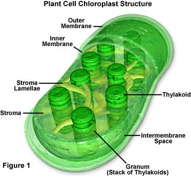
The outer, inter, and inner membrane protect the cell and also transport things like carbohydrates.
The stroma contains ribosomes, DNA, and is the location of biochemical synthesis. The stroma lamellae transports nutrients to the thylakoids. The thylakoids are the site of photosynthesis.
In photosynthesis, a light reaction occurs in the thylakoids, converting the light energy into ATP. The stroma then uses the ATP energy to convert CO2 into sugar. It does this by producing 2 glyceraldehyde 3-phosphate and combing them into a glucose molecule.
Cellular Respiration
Mitochondria
.svg/400px-Animal_mitochondrion_diagram_en_(edit).svg.png)
The outer membrane, intermembrane space, inner membrane all let proteins in and out of the cell and transport them across the membrane. The inner membrane can produce ATP. The cristae enhances the inner membrane's ability to produce ATP. The matrix helps the inner membrane make ATP using ATP synthase particles. It also contains hundreds of enzymes and 2/3 of the organelle's protein. Mitochondria have their own DNA and can make their own RNA. It also has its own Ribosomes.
Cellular Respiration is the process in which the cell converts energy from nutrients into ATP and releases waste. The mitochondria oxidizes glucose, pyruvate, and NADH in the matrix to produce ATP.
The outer, inter, and inner membrane protect the cell and also transport things like carbohydrates.
The stroma contains ribosomes, DNA, and is the location of biochemical synthesis. The stroma lamellae transports nutrients to the thylakoids. The thylakoids are the site of photosynthesis.
In photosynthesis, a light reaction occurs in the thylakoids, converting the light energy into ATP. The stroma then uses the ATP energy to convert CO2 into sugar. It does this by producing 2 glyceraldehyde 3-phosphate and combing them into a glucose molecule.
Cellular Respiration
Mitochondria
The outer membrane, intermembrane space, inner membrane all let proteins in and out of the cell and transport them across the membrane. The inner membrane can produce ATP. The cristae enhances the inner membrane's ability to produce ATP. The matrix helps the inner membrane make ATP using ATP synthase particles. It also contains hundreds of enzymes and 2/3 of the organelle's protein. Mitochondria have their own DNA and can make their own RNA. It also has its own Ribosomes.
Cellular Respiration is the process in which the cell converts energy from nutrients into ATP and releases waste. The mitochondria oxidizes glucose, pyruvate, and NADH in the matrix to produce ATP.
Subscribe to:
Posts (Atom)